
www.medical-devices.tech
31
'26
Written on Modified on
Philips and Imricor launch cardiac interventional MR lab solution
The solution combines Philips' high-performance 1.5T MRI platform and advanced interventional workflow capabilities with Imricor's exclusive portfolio of systems and consumables for MR-guided cardiac interventions.
www.philips.com
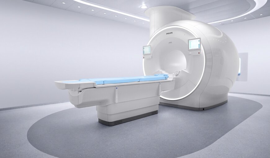
Royal Philips and Imricor Medical Systems announced the commercial release of their integrated iMR lab solution. The turnkey system pairs Philips’ 1.5-Tesla MRI infrastructure with Imricor's specialized electrophysiology (EP) devices, establishing an MR-guided cardiac intervention workflow designed to replace ionizing radiation from X-ray fluoroscopy with high-resolution soft-tissue imaging.
Real-Time Soft-Tissue Guidance and Hardware Integration
Traditional catheter ablation techniques for cardiac arrhythmias rely predominantly on X-ray fluoroscopy, which provides limited indirect visualization of soft cardiac tissue while exposing patients and medical staff to ionizing radiation. The integrated iMR solution brings advanced magnetic resonance imaging directly into the interventional suite, allowing electrophysiologists to visualize arrhythmogenic substrates, perform electroanatomical mapping, actively track catheters, and assess myocardial lesion formation in real time.
The commercial system integrates:
- Philips Cardiac MR Suite: Features SmartHeart artificial intelligence-driven scan planning alongside dedicated cardiac pulse sequences and real-time visualization applications.
- Imricor NorthStar System: Provides real-time 3D electroanatomical mapping and device tracking within the active MR imaging volume.
- Imricor Advantage-MR EP Recorder/Stimulator: Manages intracardiac electrogram acquisition, pacing stimulation, and signal filtering in the presence of strong radiofrequency and gradient fields.
- Vision-MR Catheter Family: MR-compatible ablation and diagnostic electrophysiology catheters engineered for safe operation inside the bore.
- BlueSeal Magnet Technology: Incorporates Philips' fully sealed, helium-free magnet architecture requiring only seven liters of liquid helium, reducing installation complexity and permitting placement adjacent to standard cardiology suites.
Availability and Regulatory Scope
The joint iMR lab solution is being demonstrated at the European Society of Cardiology (ESC) Congress 2026 in Munich. The platform is commercially available across CE-marked European markets and in select configurations within the United States, with expanded rollouts pending regional regulatory clearances.
Additional Context
This section details technical specifications not included in the original news release.
Performing electrophysiology interventions within a 1.5T magnetic resonance environment requires specialized radiofrequency (RF) filtering and non-ferromagnetic materials to eliminate antenna-effect heating. Standard metallic braided catheters act as resonant antennas under the 64 MHz Larmor frequency of 1.5T systems, causing induced RF currents that lead to thermal tissue injury at the catheter tip. MR-conditional catheters mitigate this risk by utilizing non-magnetic micro-coaxial wiring, ceramic internal cores, and integrated miniature RF filtering chokes or transformers that segment conductive paths, maintaining catheter tip heating below 1 degree Celsius during continuous imaging sequences.
Intracardiac electrogram (EGM) signal acquisition under active MRI requires adaptive filtering architectures to eliminate severe electromagnetic noise generated by switched magnetic field gradients and RF transmit pulses. The recording instrumentation incorporates hardware-based optoelectronic isolation barriers, optical signal transmission lines, and digital notch filters to isolate minute microvolt-level cardiac depolarization potentials from gradient-induced voltages exceeding several volts. Additionally, real-time prospective motion correction algorithms track respiratory diaphragmatic excursions and cardiac wall motion, adjusting imaging slice positions dynamically to maintain sub-millimeter anatomical registration during radiofrequency energy delivery.
Edited by Romila DSilva, Induportals Editor, with AI assistance.
The joint iMR lab solution is being demonstrated at the European Society of Cardiology (ESC) Congress 2026 in Munich. The platform is commercially available across CE-marked European markets and in select configurations within the United States, with expanded rollouts pending regional regulatory clearances.
Additional Context
This section details technical specifications not included in the original news release.
Performing electrophysiology interventions within a 1.5T magnetic resonance environment requires specialized radiofrequency (RF) filtering and non-ferromagnetic materials to eliminate antenna-effect heating. Standard metallic braided catheters act as resonant antennas under the 64 MHz Larmor frequency of 1.5T systems, causing induced RF currents that lead to thermal tissue injury at the catheter tip. MR-conditional catheters mitigate this risk by utilizing non-magnetic micro-coaxial wiring, ceramic internal cores, and integrated miniature RF filtering chokes or transformers that segment conductive paths, maintaining catheter tip heating below 1 degree Celsius during continuous imaging sequences.
Intracardiac electrogram (EGM) signal acquisition under active MRI requires adaptive filtering architectures to eliminate severe electromagnetic noise generated by switched magnetic field gradients and RF transmit pulses. The recording instrumentation incorporates hardware-based optoelectronic isolation barriers, optical signal transmission lines, and digital notch filters to isolate minute microvolt-level cardiac depolarization potentials from gradient-induced voltages exceeding several volts. Additionally, real-time prospective motion correction algorithms track respiratory diaphragmatic excursions and cardiac wall motion, adjusting imaging slice positions dynamically to maintain sub-millimeter anatomical registration during radiofrequency energy delivery.
Edited by Romila DSilva, Induportals Editor, with AI assistance.

