
www.medical-devices.tech
02
'26
Written on Modified on
Integrating CT Planning into Live Fluoroscopy Workflows
Philips UniGuide 3D Live Guidance links pre-procedural computed tomography data directly to the Philips Azurion image-guided therapy system for real-time procedural guidance.
www.philips.com
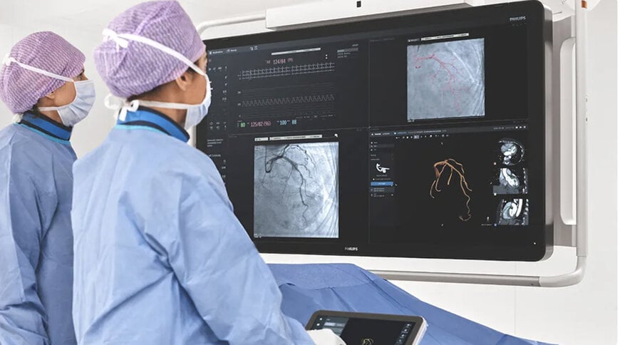
Minimally invasive catheterization laboratories face a recurring operational bottleneck: high-resolution anatomical datasets generated prior to an intervention remain trapped in isolated diagnostic workstations. In percutaneous coronary interventions (PCI), structural heart operations, and complex endovascular repairs, operators frequently rely on coronary computed tomography angiography (CCTA) to evaluate calcium distribution, vessel morphology, and plaque topography. Translating these static pre-operative three-dimensional cross-sections to dynamic two-dimensional X-ray fluoroscopy traditionally demands continuous mental spatial co-registration, repeatedly diverting operator focus away from the primary patient field.
Operating Architecture and Cath Lab Hardware Integration
The platform addresses this intra-procedural discontinuity by directly embedding pre-procedural volumetric models into the sterile catheterization field. Utilizing the Azurion system architecture, the technology connects to datasets via the Philips Advanced Visualization Workspace as well as validated, open-interface third-party planning packages.
During live catheter navigation, volumetric models display on the central FlexVision monitor through calibrated three-dimensional digital overlays or synchronized side-by-side configurations. Rather than requiring secondary control room interventions, the operating cardiologist manipulates volumetric projections using the sterile tableside Touch Screen Module (TSM). Motorized C-arm gantry movements align automatically with the 3D CT volume rendering, ensuring the fluoroscopic projection angle matches target anatomical views without manual geometric recalibration.
Percutaneous Coronary and Vascular Applications
In coronary interventions, accessing synchronized CCTA datasets during active wiring provides live spatial markers for vessel landing zones and plaque composition. Interventional cardiologists can reference vessel dimensions, bifurcation geometry, and vessel wall calcium load alongside live radiopaque guidewire progression.
According to Prof. Javier Escaned, MD, PhD, Head of the Interventional Cardiology Section at Hospital Clínico San Carlos and Complutense University in Madrid:
"Access to the CT angiogram allows us to assess relevant aspects of the vessels we are going to treat, improve procedural planning, and improve the precision of the intervention. We can also review specific aspects of the CT inside the cathlab using the table-side touchscreen (TSM) and synchronize the movement of the 3D CT rendering with the C-arm. Together, these capabilities contribute to better guidance and higher precision of the interventions."
Usability Evaluation and Regulatory Status
In an internal manufacturer claims validation study comprising 15 clinical operators, 93 percent of participants reported that integrating diagnostic planning into the procedural flow improves catheterization suite operational efficiency, while 80 percent stated that procedural automation aids physician focus during interventions.
The medical software achieved European CE mark conformity and entered commercial distribution across CE-accepting territories on October 23, 2026. The product remains outside commercial distribution within the United States pending relevant regulatory clearance pathways.
Additional Context:
This section details technical specifications and competitive benchmarking not included in the original product announcement.
The technological shift toward intra-procedural multimodality image fusion reflects established clinical trends across interventional cardiology and endovascular surgery. Standard fluoroscopy relies solely on two-dimensional radiopaque projections and repeated iodinated contrast injections. By contrast, three-dimensional fusion navigation systems register volumetric imaging—such as contrast-enhanced CT or rotational cone-beam CT (CBCT)—onto the real-time fluoroscopic detector coordinates. This established approach reduces total fluoroscopy exposure time and overall nephrotoxic contrast media usage during complex interventions, such as chronic total occlusion (CTO) crossing and transcatheter aortic valve replacement (TAVR).
Within the competitive interventional imaging sector, comparable multimodality fusion solutions operate on similar geometric co-registration principles:
Siemens Healthineers syngo Fusion Package: Deployed on the Artis zee, Artis Q, and ARTIS pheno angiography systems, this platform supports rigid 2D/3D and 3D/3D registration. It aligns pre-procedural DICOM CT or magnetic resonance datasets with live fluoroscopy or intra-procedural syngo DynaCT datasets using anatomical landmarks and rotational angiographic baselines.
GE HealthCare Valve ASSIST and CT Fusion: Operating within the Innova and Allia angiography ecosystems, this architecture co-registers pre-acquired 3D CT angiography datasets with live fluoroscopy and real-time transesophageal echocardiography (TEE). The system automatically aligns the C-arm projection angle to predetermined anatomical landmarks for structural heart procedures such as TAVR and left atrial appendage occlusion (LAAO).
While competing platforms rely primarily on dedicated proprietary workstations or specialized intra-operative 3D acquisition runs (such as DynaCT), Philips positions the UniGuide framework around open compatibility with external vendor planning software and direct touch control through the native Azurion tableside user interface.
Edited by Sucithra Mani, Induportals editor – adapted by AI.
www.philips.com

