
www.medical-devices.tech
25
'26
Written on Modified on
Four-Dimensional Magnetic Resonance Imaging for Radiotherapy Simulation
Royal Philips developed a motion-resolved imaging solution that manages respiratory artifacts to improve soft-tissue visualization, assisting clinicians in precisely targeting abdominal tumors during oncology treatment planning.
www.philips.com
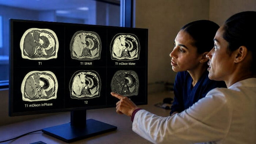
Motion during respiration presents challenges in abdominal radiotherapy planning, as breathing variations can introduce artifacts and blur structural boundaries in the liver and pancreas. Standard mitigation techniques, including breath-hold protocols or abdominal compression devices, often reduce patient comfort and limit reproducibility.
The integration of four-dimensional magnetic resonance radiotherapy simulation (4D MR-RT) addresses this by capturing anatomical data during normal, free-breathing cycles. The system utilizes SmartSpeed reconstruction technology to track the respiratory cycle and segregate data into distinct inhale and exhale phases. Clinicians can reconstruct up to 10 distinct respiratory phases, allowing for precise tracking of moving target volumes and adjacent organs at risk.
Multi-Contrast Acquisition and Workflow Integration
The imaging solution provides simultaneous multi-contrast 4D imaging, generating both T1-weighted and T2-weighted sequences within the same motion-resolved framework. These complementary contrasts offer distinct clinical utility: T1 sequences highlight anatomical boundaries and vascular structures, while T2 sequences assist in identifying fluid-rich lesions and soft-tissue pathologies. Optional fat suppression techniques are integrated to further isolate target tissues from surrounding adipose layers.
To fit standard linear accelerator workflows, the system generates specific secondary image outputs. These include individual respiratory phase datasets, mid-ventilation images, and mid-position reconstructions. These outputs provide quantitative spatial coordinates for target contouring, facilitating precise margin calculations for advanced delivery techniques like stereotactic body radiotherapy. Regulatory clearance has been granted for clinical deployment via 510(k) clearance in the United States and the CE Mark in Europe.
Additional Context: Technical Specifications and Competitive Benchmarking
Magnetic resonance simulation requires precise spatial fidelity and motion management to match the geometric accuracy of computed tomography, which remains the historical standard for radiation dose calculations.
In the market for motion-managed MR simulation, alternative approaches typically rely on external respiratory gating hardware, such as bellows belts or optical surface tracking, to synchronize image acquisition with the breathing cycle. The Philips 4D MR-RT approach leverages internal data-driven gating via SmartSpeed algorithms, reducing reliance on external hardware setups.
Competitively, Siemens Healthineers utilizes a technology known as Free-Breathing GRASP (Golden-angle Radial Sparse Parallel) on its Biograph mMR and MAGNETOM systems to handle continuous, non-gated free-breathing abdominal exams. While GRASP uses radial k-space sampling to resolve motion retrospectively into arbitrary time frames, the Philips 4D MR-RT implementation focuses on generating up to 10 discrete respiratory phases with synchronized T1 and T2 contrasts specifically optimized for radiotherapy contouring pipelines.
GE HealthCare addresses this clinical need through its Silent Scan and PROPELLER (Periodically Rotated Overlapping Parallel Lines with Enhanced Reconstruction) sequences, which mitigate motion artifacts by oversampling the center of k-space. However, standard PROPELLER sequences do not inherently sort data into a 4D time series based on respiratory phase, giving 4D MR-RT an analytical advantage in quantifying specific tumor trajectory paths for internal target volume definition.
Edited by Evgeny Churilov, Induportals Media - Adapted by AI.
www.philips.com

