
www.medical-devices.tech
23
'26
Written on Modified on
GE HealthCare announces five new global clinical collaborations
Leading institutions will evaluate StarGuide GX across quantitative imaging, dynamic imaging and emerging theranostic applications.
www.gehealthcare.com
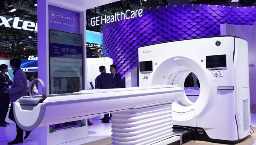
GE HealthCare has initiated a global multicenter evaluation program partnering with five clinical and research institutions across North America, Europe, the Middle East, and Asia to assess its StarGuide GX digital 4D SPECT/CT system. The research initiative targets molecular imaging environments, focusing on precision quantitative verification, dynamic tracer kinetics, and image reconstruction performance in routine oncology and emerging theranostic procedures.
Clinical Demands and Molecular Imaging Precision
Nuclear medicine workflows increasingly require absolute biological quantification to verify therapeutic response, guide personalized dosing, and support targeted radionuclide therapy. Traditional analog single-photon emission computed tomography systems often face sensitivity limitations, prolonged acquisition times, and mechanical restrictions when imaging high-energy or low-abundance isotopes. To validate advanced detector mechanics under diverse clinical regimens, GE HealthCare is collaborating with Hadassah Medical Center, Mayo Clinic, Oncopole Claudius Regaud, Peking University First Hospital, and Rambam Health Care Campus to benchmark dynamic imaging protocols across varied patient demographics.
Detector Architecture and Reconstruction Hardware
The StarGuide GX system integrates dual-sided cadmium zinc telluride solid-state detector technology to achieve simultaneous high spatial resolution and volumetric sensitivity. The motorized gantry design enables individual detectors to track patient contours closely, maximizing photon capture geometry. A dual-collimator configuration eliminates manual collimator exchanges, removing mechanical swap latency between low-energy and medium-energy imaging protocols.
The system incorporates Low-Medium Energy collimation engineered to provide up to 2.67-times higher system volume sensitivity compared to standard configurations. Compute-intensive iterative reconstruction pipelines execute on onboard graphics processors utilizing NVIDIA RTX accelerated computing, enabling fast tomographic rendering of dynamic, time-resolved data sets. The energy acceptance architecture accommodates a broad radionuclide spectrum, facilitating research into high-energy alpha-emitting isotopes such as Actinium-225 used in targeted alpha therapies.
Institutional Evaluation Scenarios and Workflow Validation
Participating centers will evaluate distinct operational and technical facets of the platform:
- Hadassah Medical Center in Israel evaluates advanced quantitative processing following a platform field upgrade.
- Mayo Clinic in the United States analyzes high-frame-rate dynamic acquisition algorithms and quantitative performance baselines.
- Oncopole Claudius Regaud at the Institut Universitaire du Cancer de Toulouse in France deploys the platform across routine diagnostic oncology workflows and high-energy theranostic clinical research.
- Peking University First Hospital in China assesses novel radiotracer tracking and preclinical imaging applications.
- Rambam Health Care Campus in Israel monitors clinical throughput, ergonomics, and daily workflow consistency across an extensive portfolio of nuclear medicine tracers.
Operational Impact on Theranostic Workflows
Transitioning to solid-state digital detection combined with accelerated reconstruction reduces scan acquisition duration and improves lesion detectability. Automating physical collimator transitions streamlines technical workflows, lowers occupational radiation handling risks, and delivers repeatable quantitative measurement metrics essential for continuous therapeutic monitoring in molecular oncology.
Edited by Romila DSilva, Induportals Editor, with AI assistance.

