
www.medical-devices.tech
06
'26
Written on Modified on
Fraunhofer IGD develops AI software for early osteoporosis detection
BMDNow analyzes existing CT scans to calculate vertebral bone density, identifying osteoporosis and osteopenia without additional specialized examinations.
www.fraunhofer.de
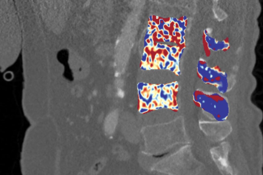
© Fraunhofer IGD: Cross section of the spine with a color-coded density map. Trabecular bone density decreases from blue through yellow to red. The scale ranges from red (0 mg/mm³) to blue (800 mg/mm³).
Asymptomatic skeletal degradation presents a major clinical challenge because significant bone loss frequently remains unnoticed until a fragility fracture occurs. To address this diagnostic gap in clinical radiology, researchers at the Fraunhofer Institute for Computer Graphics Research IGD, in partnership with Goethe University Frankfurt and garritz online media international, developed the BMDNow software to extract opportunistic bone mineral density measurements directly from existing computed tomography scans.
Diagnostic Workflow Integration and Opportunistic Screening
Clinical diagnosis of skeletal conditions such as osteopenia and osteoporosis primarily depends on dual-energy X-ray absorptiometry. Dual-energy X-ray absorptiometry operates as an isolated procedure, typically ordered only after a patient exhibits clinical symptoms, back pain, or mechanical bone failure. This sequential diagnostic pathway leads to late intervention and imposes cumulative radiation doses on at-risk demographics.
The BMDNow framework integrates directly into digital radiological workflows by utilizing pre-existing routine volumetric data, including routine thoracic, abdominal, or cardiac computed tomography examinations. By processing scans performed for unrelated clinical indications, the automated tool calculates volumetric bone mineral density as a secondary analytical output. This workflow reduces overall radiation burden and optimizes radiologist review times by avoiding dedicated follow-up examinations.
Neural Network Segmentation and Calibration Mechanics
The computational architecture of the software combines neural network algorithms with analytical imaging techniques to process volumetric DICOM data. The process focuses on the spongy trabecular compartment of individual vertebral bodies, which exhibits early metabolic turnover and structural degradation during systemic bone loss.
During execution, neural networks segment the trabecular boundaries of targeted vertebrae from the denser cortical shell. The system evaluates dual-energy computed tomography attenuation properties across different X-ray energy spectra. This separation enables material decomposition to quantify specific biological fractions, principally hydroxyapatite calcium mineral content and marrow adipose tissue.
A three-dimensional mapping engine computes local tissue density distribution and applies pseudocolor visualization to represent spatial variations. Quantitative volumetric bone mineral density values are derived using empirical calibration curves generated from physical reference phantom models. These calibrated values are evaluated against diagnostic thresholds to distinguish between physiological bone mass, osteopenia, and advanced osteoporosis.
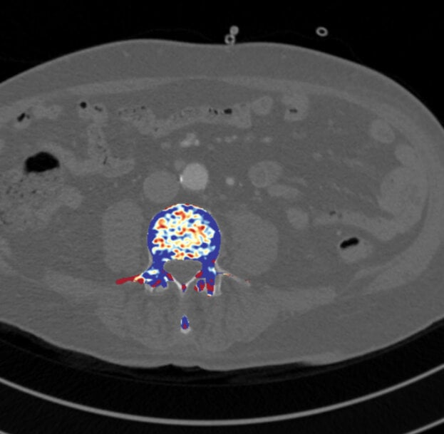
© Fraunhofer IGD: Cross-section of a vertebra with color-coded density map
Algorithm Validation and Multi-Energy Expansion
Model training and validation utilized patient imaging cohorts supplied by Frankfurt University Hospital to ensure analytical reliability across clinical variability. While initial system deployment operates on dual-energy computed tomography due to its intrinsic material decomposition capabilities, the consortium is expanding the neural network architecture to process standard single-energy computed tomography volumes. This transition requires algorithmic calibration adjustments to account for scanner-specific tube voltages and beam hardening artifacts without dual-spectrum acquisition.
The project consortium received second place in the Hessen Ideen competition in 2025, supported by funding from Hessen Agentur. System development focuses on compiling an expanded reference database to improve algorithm generalizability across diverse multi-vendor imaging platforms. Software prototyping and clinical validation remain in progress, with regulatory submissions for medical device certification planned within a one- to two-year development horizon.
Additional Context:
This section details technical specifications and competitive benchmarking not included in the original product announcement.
Opportunistic quantitative computed tomography, often designated as QCT or asynchronous QCT, represents an active domain in medical imaging informatics aimed at identifying metabolic bone disease without dedicated phantom scanning during each patient acquisition. In traditional quantitative computed tomography workflows, simultaneous scanning of a solid calibration phantom beneath the patient was required to convert Hounsfield Units into volumetric bone mineral density equivalents expressed in milligrams of hydroxyapatite per cubic centimeter (mg/cm³). Modern deep learning approaches substitute physical phantoms by implementing internal tissue calibration, using blood pool attenuation or muscular tissue references to normalize inter-scanner variability.
Standard dual-energy X-ray absorptiometry measures areal bone mineral density (g/cm²), which represents a two-dimensional projection susceptible to structural artifacts such as aortic calcifications, spinal degenerative changes, and osteophytes. In contrast, three-dimensional quantitative computed tomography isolates trabecular bone in the vertebral centrum, providing higher sensitivity to early metabolic changes. American College of Radiology guidelines define volumetric trabecular bone mineral density thresholds measured by quantitative computed tomography as greater than 120 mg/cm³ for normal bone, 80 to 120 mg/cm³ for osteopenia, and below 80 mg/cm³ for osteoporosis.
Commercial artificial intelligence solutions addressing opportunistic osteoporosis screening include systems such as Zebra Medical Vision (Nanox.AI) HealthOST and ImageBiopsy Lab's musculoskeletal assessment tools, which are clearance-evaluated by regulatory agencies under radiological computer-assisted diagnostic software categories. These commercial implementations primarily process single-energy computed tomography series through automatic region-of-interest segmentation and asynchronous density mapping. The application of dual-energy computed tomography, as explored in BMDNow, enhances analytical specificity by directly separating virtual non-calcium and calcium-specific attenuation profiles, mitigating errors caused by variable yellow and red marrow conversion that typically confound single-energy attenuation values.
Edited by Natania Lyngdoh, Induportals editor, with AI assistance.
www.fraunhofer.com
Asymptomatic skeletal degradation presents a major clinical challenge because significant bone loss frequently remains unnoticed until a fragility fracture occurs. To address this diagnostic gap in clinical radiology, researchers at the Fraunhofer Institute for Computer Graphics Research IGD, in partnership with Goethe University Frankfurt and garritz online media international, developed the BMDNow software to extract opportunistic bone mineral density measurements directly from existing computed tomography scans.
Diagnostic Workflow Integration and Opportunistic Screening
Clinical diagnosis of skeletal conditions such as osteopenia and osteoporosis primarily depends on dual-energy X-ray absorptiometry. Dual-energy X-ray absorptiometry operates as an isolated procedure, typically ordered only after a patient exhibits clinical symptoms, back pain, or mechanical bone failure. This sequential diagnostic pathway leads to late intervention and imposes cumulative radiation doses on at-risk demographics.
The BMDNow framework integrates directly into digital radiological workflows by utilizing pre-existing routine volumetric data, including routine thoracic, abdominal, or cardiac computed tomography examinations. By processing scans performed for unrelated clinical indications, the automated tool calculates volumetric bone mineral density as a secondary analytical output. This workflow reduces overall radiation burden and optimizes radiologist review times by avoiding dedicated follow-up examinations.
Neural Network Segmentation and Calibration Mechanics
The computational architecture of the software combines neural network algorithms with analytical imaging techniques to process volumetric DICOM data. The process focuses on the spongy trabecular compartment of individual vertebral bodies, which exhibits early metabolic turnover and structural degradation during systemic bone loss.
During execution, neural networks segment the trabecular boundaries of targeted vertebrae from the denser cortical shell. The system evaluates dual-energy computed tomography attenuation properties across different X-ray energy spectra. This separation enables material decomposition to quantify specific biological fractions, principally hydroxyapatite calcium mineral content and marrow adipose tissue.
A three-dimensional mapping engine computes local tissue density distribution and applies pseudocolor visualization to represent spatial variations. Quantitative volumetric bone mineral density values are derived using empirical calibration curves generated from physical reference phantom models. These calibrated values are evaluated against diagnostic thresholds to distinguish between physiological bone mass, osteopenia, and advanced osteoporosis.
© Fraunhofer IGD: Cross-section of a vertebra with color-coded density map
Algorithm Validation and Multi-Energy Expansion
Model training and validation utilized patient imaging cohorts supplied by Frankfurt University Hospital to ensure analytical reliability across clinical variability. While initial system deployment operates on dual-energy computed tomography due to its intrinsic material decomposition capabilities, the consortium is expanding the neural network architecture to process standard single-energy computed tomography volumes. This transition requires algorithmic calibration adjustments to account for scanner-specific tube voltages and beam hardening artifacts without dual-spectrum acquisition.
The project consortium received second place in the Hessen Ideen competition in 2025, supported by funding from Hessen Agentur. System development focuses on compiling an expanded reference database to improve algorithm generalizability across diverse multi-vendor imaging platforms. Software prototyping and clinical validation remain in progress, with regulatory submissions for medical device certification planned within a one- to two-year development horizon.
Additional Context:
This section details technical specifications and competitive benchmarking not included in the original product announcement.
Opportunistic quantitative computed tomography, often designated as QCT or asynchronous QCT, represents an active domain in medical imaging informatics aimed at identifying metabolic bone disease without dedicated phantom scanning during each patient acquisition. In traditional quantitative computed tomography workflows, simultaneous scanning of a solid calibration phantom beneath the patient was required to convert Hounsfield Units into volumetric bone mineral density equivalents expressed in milligrams of hydroxyapatite per cubic centimeter (mg/cm³). Modern deep learning approaches substitute physical phantoms by implementing internal tissue calibration, using blood pool attenuation or muscular tissue references to normalize inter-scanner variability.
Standard dual-energy X-ray absorptiometry measures areal bone mineral density (g/cm²), which represents a two-dimensional projection susceptible to structural artifacts such as aortic calcifications, spinal degenerative changes, and osteophytes. In contrast, three-dimensional quantitative computed tomography isolates trabecular bone in the vertebral centrum, providing higher sensitivity to early metabolic changes. American College of Radiology guidelines define volumetric trabecular bone mineral density thresholds measured by quantitative computed tomography as greater than 120 mg/cm³ for normal bone, 80 to 120 mg/cm³ for osteopenia, and below 80 mg/cm³ for osteoporosis.
Commercial artificial intelligence solutions addressing opportunistic osteoporosis screening include systems such as Zebra Medical Vision (Nanox.AI) HealthOST and ImageBiopsy Lab's musculoskeletal assessment tools, which are clearance-evaluated by regulatory agencies under radiological computer-assisted diagnostic software categories. These commercial implementations primarily process single-energy computed tomography series through automatic region-of-interest segmentation and asynchronous density mapping. The application of dual-energy computed tomography, as explored in BMDNow, enhances analytical specificity by directly separating virtual non-calcium and calcium-specific attenuation profiles, mitigating errors caused by variable yellow and red marrow conversion that typically confound single-energy attenuation values.
Edited by Natania Lyngdoh, Induportals editor, with AI assistance.
www.fraunhofer.com

