
www.medical-devices.tech
02
'26
Written on Modified on
Fraunhofer develops Sustainable Printing Inks for Functional Organs
PhysioINK uses collagen, elastin and cellulose derivatives to create printable bioinks for tissue models, drug development and future regenerative medicine.
www.fraunhofer.de
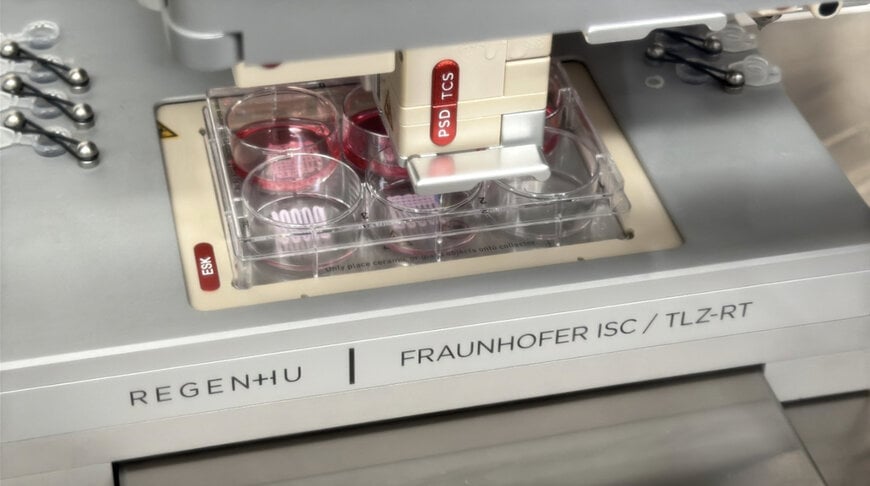
© K. Selsam / Fraunhofer ISC: 3D printing freestanding lattice structures
Fabricating transplantable organ constructs and predictive pharmacological tissue assays faces a material-science bottleneck: conventional bioinks either lack printability in their native states or require chemical modifications that disrupt physiological cellular interactions. To overcome this limitation, a research consortium led by the Fraunhofer Institute for Silicate Research ISC, in collaboration with Fraunhofer IAP, Fraunhofer IMWS, and Fraunhofer IZI-BB, is developing unmodified protein-based bioinks within the PhysioINK project to replicate native extracellular matrix architectures for drug screening and regenerative implants.
Macromolecular Stabilization and Self-Assembly Mechanism
Extracellular matrices in mammalian tissues consist predominantly of fibrous proteins, notably type I collagen, type IV collagen, and elastin, which account for 60% to 100% of soft-tissue dry mass. Under physiological pH and temperature, native collagen spontaneously self-assembles into insolubilized, cross-linked fibrillar networks. In conventional 3D bioprinting systems, this premature gelation either causes continuous shear clogging in extrusion nozzles or forces researchers to chemically modify the backbone with methacrylates, altering cellular binding sites.
The PhysioINK project circumvents chemical alteration by stabilizing native proteins using electrostatic shielding. Unmodified collagen and elastin molecules display net positive surface charges in neutral aqueous solutions. The research team synthesizes customized, negatively charged cellulose sulfates (CS) whose molecular geometry and charge distribution match the protein backbone. These polysaccharide chains encapsulate the cationic domains of the proteins, suppressing spontaneous fibrillogenesis and preserving a fluid, printable state at neutral pH.
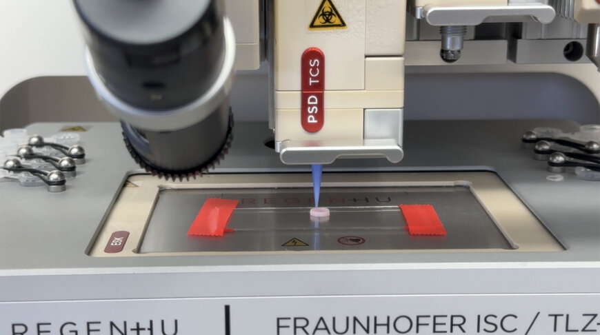
© K. Selsam / Fraunhofer ISC: 3D printing free-standing ring structures
Following extrusion through dispensing nozzles, deposition onto the target substrate triggers controlled self-assembly via a thermal mechanism. Raising the ambient platform temperature disrupts the temporary polysaccharide-protein coordination complex, allowing the native polypeptide chains to self-organize into natural fibrous and network-like architectures. This thermal release enables the formation of physiological fibrillar structures without the toxic photoinitiators or chemical cross-linkers required by synthetic hydrogels.
Print Rheology and Extrusion Optimization
Systematic printability assessments evaluating type I collagen inks at concentrations of 3%, 5%, and 7% revealed direct correlations between mass concentration, shear thinning, and post-deposition shape fidelity. While 3% formulations experienced lateral slump and dimensional spreading prior to thermal gelation, inks formulated at 5% and 7% successfully maintained shape integrity to form freestanding, high-aspect-ratio geometries across multi-axis printer platforms.
To assess the operational viability of these bioinks in distributed medical research pipelines, the consortium conducted transport and shelf-life qualification trials. The formulated type I collagen inks remained in an ungelled, fully soluble state for up to nine months when stored at 4°C, and sustained freeze-thaw cycles down to -20°C without degradation of subsequent thermal gelation kinetics. Thermal logging of inter-laboratory refrigerated transit tests confirmed that fluid printability is fully preserved after 48 hours at temperatures up to 8.0°C.
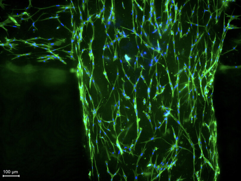
© Tobias Weigel/Fraunhofer ISC: A microscope reveals the living tissue cells (fibroblasts) in the printed grid structure: Cell nuclei are blue and the cytoskeleton is green.
Preclinical Model Validation and Clinical Scope
The project targets two primary tissue paradigms scheduled across a development roadmap running from February 2025 through January 2028: standardized intestinal tumor geometries for high-throughput oncology therapeutics screening, and functional cardiac patches for myocardial repair. In preliminary multi-layer constructs, the consortium produced vascularized skin models, integrating real-time metabolic sensor suites to measure cellular oxygen consumption rates and nutrient perfusion within the newly extruded scaffolds.
Project coordinator Tobias Weigel of Fraunhofer ISC indicated that moving away from synthetic polymers to native, unmodified physiological proteins provides the specific microstructural cues required to replicate authentic human organ behavior.
Additional Context:
This section details technical specifications and competitive benchmarking not included in the original product announcement.
Conventional 3D bioprinting hydrogels rely heavily on gelatin methacryloyl (GelMA), a semi-synthetic biomaterial produced by functionalizing porcine or bovine gelatin with methacrylic anhydride. While GelMA supports rapid free-radical polymerization under ultraviolet or blue-light exposure (365 nm to 405 nm) utilizing photoinitiators like lithium phenyl-2,4,6-trimethylbenzoylphosphinate (LAP), the residual covalent cross-links deviate structurally from natural fibrillar collagen networks. These synthetic chemical bonds limit cell-mediated matrix metalloproteinase (MMP) remodeling and run the risk of reactive oxygen species cytotoxicity during photocuring.
Standard unmodified commercial collagen bioinks typically feature low dynamic viscosities and slow gelation kinetics, demanding support-bath printing methods such as Freeform Reversible Embedding of Suspended Hydrogels (FRESH) to prevent structural collapse of low-viscosity (under 0.5% weight-per-volume) solutions. In contrast, the cellulose-stabilized 5% and 7% collagen formulations within PhysioINK achieve standalone yield-stress behavior, enabling direct extrusion on standard bioprinting systems without secondary microgel support baths.
Academic and clinical research initiatives utilizing human-derived matrices increasingly benchmark cellular responses against ISO 10993 biological evaluation of medical devices and ASTM F2150 standards for biomaterial tissue engineering scaffolds. Eliminating xenogeneic animal components and chemical adulterants aligns tissue engineering directly with European Medicines Agency (EMA) advanced therapy medicinal product (ATMP) regulatory guidelines, removing immunogenic degradation vectors during long-term clinical implantation.
Edited by Natania Lyngdoh, Induportals editor, with AI assistance.
www.fraunhofer.com
Fabricating transplantable organ constructs and predictive pharmacological tissue assays faces a material-science bottleneck: conventional bioinks either lack printability in their native states or require chemical modifications that disrupt physiological cellular interactions. To overcome this limitation, a research consortium led by the Fraunhofer Institute for Silicate Research ISC, in collaboration with Fraunhofer IAP, Fraunhofer IMWS, and Fraunhofer IZI-BB, is developing unmodified protein-based bioinks within the PhysioINK project to replicate native extracellular matrix architectures for drug screening and regenerative implants.
Macromolecular Stabilization and Self-Assembly Mechanism
Extracellular matrices in mammalian tissues consist predominantly of fibrous proteins, notably type I collagen, type IV collagen, and elastin, which account for 60% to 100% of soft-tissue dry mass. Under physiological pH and temperature, native collagen spontaneously self-assembles into insolubilized, cross-linked fibrillar networks. In conventional 3D bioprinting systems, this premature gelation either causes continuous shear clogging in extrusion nozzles or forces researchers to chemically modify the backbone with methacrylates, altering cellular binding sites.
The PhysioINK project circumvents chemical alteration by stabilizing native proteins using electrostatic shielding. Unmodified collagen and elastin molecules display net positive surface charges in neutral aqueous solutions. The research team synthesizes customized, negatively charged cellulose sulfates (CS) whose molecular geometry and charge distribution match the protein backbone. These polysaccharide chains encapsulate the cationic domains of the proteins, suppressing spontaneous fibrillogenesis and preserving a fluid, printable state at neutral pH.
© K. Selsam / Fraunhofer ISC: 3D printing free-standing ring structures
Following extrusion through dispensing nozzles, deposition onto the target substrate triggers controlled self-assembly via a thermal mechanism. Raising the ambient platform temperature disrupts the temporary polysaccharide-protein coordination complex, allowing the native polypeptide chains to self-organize into natural fibrous and network-like architectures. This thermal release enables the formation of physiological fibrillar structures without the toxic photoinitiators or chemical cross-linkers required by synthetic hydrogels.
Print Rheology and Extrusion Optimization
Systematic printability assessments evaluating type I collagen inks at concentrations of 3%, 5%, and 7% revealed direct correlations between mass concentration, shear thinning, and post-deposition shape fidelity. While 3% formulations experienced lateral slump and dimensional spreading prior to thermal gelation, inks formulated at 5% and 7% successfully maintained shape integrity to form freestanding, high-aspect-ratio geometries across multi-axis printer platforms.
To assess the operational viability of these bioinks in distributed medical research pipelines, the consortium conducted transport and shelf-life qualification trials. The formulated type I collagen inks remained in an ungelled, fully soluble state for up to nine months when stored at 4°C, and sustained freeze-thaw cycles down to -20°C without degradation of subsequent thermal gelation kinetics. Thermal logging of inter-laboratory refrigerated transit tests confirmed that fluid printability is fully preserved after 48 hours at temperatures up to 8.0°C.
© Tobias Weigel/Fraunhofer ISC: A microscope reveals the living tissue cells (fibroblasts) in the printed grid structure: Cell nuclei are blue and the cytoskeleton is green.
Preclinical Model Validation and Clinical Scope
The project targets two primary tissue paradigms scheduled across a development roadmap running from February 2025 through January 2028: standardized intestinal tumor geometries for high-throughput oncology therapeutics screening, and functional cardiac patches for myocardial repair. In preliminary multi-layer constructs, the consortium produced vascularized skin models, integrating real-time metabolic sensor suites to measure cellular oxygen consumption rates and nutrient perfusion within the newly extruded scaffolds.
Project coordinator Tobias Weigel of Fraunhofer ISC indicated that moving away from synthetic polymers to native, unmodified physiological proteins provides the specific microstructural cues required to replicate authentic human organ behavior.
Additional Context:
This section details technical specifications and competitive benchmarking not included in the original product announcement.
Conventional 3D bioprinting hydrogels rely heavily on gelatin methacryloyl (GelMA), a semi-synthetic biomaterial produced by functionalizing porcine or bovine gelatin with methacrylic anhydride. While GelMA supports rapid free-radical polymerization under ultraviolet or blue-light exposure (365 nm to 405 nm) utilizing photoinitiators like lithium phenyl-2,4,6-trimethylbenzoylphosphinate (LAP), the residual covalent cross-links deviate structurally from natural fibrillar collagen networks. These synthetic chemical bonds limit cell-mediated matrix metalloproteinase (MMP) remodeling and run the risk of reactive oxygen species cytotoxicity during photocuring.
Standard unmodified commercial collagen bioinks typically feature low dynamic viscosities and slow gelation kinetics, demanding support-bath printing methods such as Freeform Reversible Embedding of Suspended Hydrogels (FRESH) to prevent structural collapse of low-viscosity (under 0.5% weight-per-volume) solutions. In contrast, the cellulose-stabilized 5% and 7% collagen formulations within PhysioINK achieve standalone yield-stress behavior, enabling direct extrusion on standard bioprinting systems without secondary microgel support baths.
Academic and clinical research initiatives utilizing human-derived matrices increasingly benchmark cellular responses against ISO 10993 biological evaluation of medical devices and ASTM F2150 standards for biomaterial tissue engineering scaffolds. Eliminating xenogeneic animal components and chemical adulterants aligns tissue engineering directly with European Medicines Agency (EMA) advanced therapy medicinal product (ATMP) regulatory guidelines, removing immunogenic degradation vectors during long-term clinical implantation.
Edited by Natania Lyngdoh, Induportals editor, with AI assistance.
www.fraunhofer.com

