

www.medical-devices.tech
28
'26
Written on
Modified on
ZEISS News
ZEISS Presents Retinal Diagnostic And Surgical Innovations At EURETINA 2026
Integrated diagnostic imaging, head-mounted surgical visualization, and micro-incisional vitrectomy instruments unify the clinical pathway from disease screening to posterior segment surgery.
www.zeiss.com
Carl Zeiss Meditec AG presented an expanded retinal care portfolio at the 2026 European Society of Retina Specialists congress in Vienna, held from October 1 through October 4. The developments address vitreoretinal surgery, clinical diagnostics, and surgical visualization across ophthalmology practices and ambulatory surgical centers.
Multimodal Diagnostic Imaging Architecture
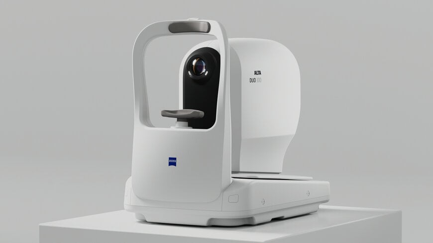
Diagnostic additions introduce the ZEISS ALTA DUO 300, a compact multimodal imager designed to combine optical coherence tomography and fundus photography within a single hardware footprint. The automated platform conducts non-mydriatic structural scanning and color photographic documentation via unified optical alignment, reducing operator dependency and patient transit times between separate testing instruments. The system expands the clinical range alongside established standalone diagnostic platforms, specifically CIRRUS optical coherence tomography and CLARUS widefield fundus imaging systems.
Head-Mounted Digital Visualization and Ergonomic Integration
Surgical visualization capabilities expand through the integration of the HMDmd CR3 head-mounted three-dimensional display with the ZEISS ARTEVO 850 digital surgical microscope. The wearable display system provides surgeons with direct stereoscopic heads-up visualization without relying on stationary three-dimensional monitors. The hardware architecture transmits digital microscopy video feeds directly to the operator alongside intraoperative overlays, including depth of field controls, digital color adjustments, and real-time exposure feedback, maintaining full visualization while supporting neutral postural alignment during complex surgery.
Vitreoretinal Surgical Dyes and Cutting Mechanics
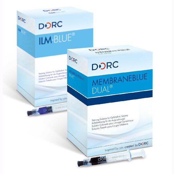
Vitreoretinal surgical tools highlight the milestone of three million cumulative clinical procedures using selective membrane stains, specifically ILM-BLUE and MEMBRANEBLUE-DUAL. Both dyes incorporate a four percent polyethylene glycol carrier solution that increases solution density relative to balanced salt solution. When injected into the vitreous cavity, the dye forms a cohesive droplet that settles directly onto the retinal surface, staining target internal limiting membranes and epiretinal membranes without diffuse intravitreal dispersion.
Mechanical instrumentation features the 27-gauge TDC VELOCE two-dimensional cutter. The cutter incorporates a dual-edge blade geometry that severs tissue during both forward and backward cycles, effectively doubling cutting frequency relative to single-action cutters. Clinical data from 27-gauge vitrectomy procedures indicate that the continuous duty cycle and increased aspiration flow rate shorten core vitrectomy procedure duration by up to 41 percent compared to previous two-dimensional cutting assemblies.
Corporate Integration and Technical Presentations
The innovations reflect the operational transition of Dutch Ophthalmic Research Center under the single corporate identity of ZEISS. The brand consolidation unifies equipment procurement, service contracts, and consumable supply chains across posterior segment diagnostic and surgical technologies.
The technical program at EURETINA 2026 in Vienna included presentations across the Retina Forum and Innovation Spotlight on surgical robotics and artificial intelligence therapy, accompanied by educational symposia focused on multimodal retinal vascular angiography and vitrectomy case management.
Additional Context:
This section details technical specifications and competitive benchmarking not included in the original product announcement
Ophthalmic diagnostics and vitreoretinal surgical platforms are evaluated against standardized optical resolution, cut-speed metrics, and fluidic stability criteria. Multimodal combined systems occupy a distinct mid-tier clinic footprint evaluated by spectral-domain optical coherence tomography acquisition speeds (typically 50,000 to 100,000 axial scans per second) and fundus camera sensor resolution. Comparable platforms include the Topcon Maestro2, which pairs an automated spectral-domain scanner running at 50,000 axial scans per second with a 12-megapixel color fundus camera, and the Canon OCT-HS100 series. Higher-tier specialist units, such as the Heidelberg Engineering SPECTRALIS platform, utilize scanning laser ophthalmoscopy and achieve up to 125,000 axial scans per second with modular anterior, angiography, and autofluorescence configurations.
In vitreoretinal surgery, competitive benchmarking centers on cutter mechanics and effective cut rates to minimize retinal traction during peripheral vitreous shaving. Standard single-edge cutters on competitive platforms, such as the Alcon Constellation Vision System and Bausch + Lomb Stellaris Elite, achieve cutting rates between 7,500 and 10,000 cuts per minute using single-action pneumatic or electric actuators. DORC TDC cutters utilize a reciprocating double-sided guillotine blade, maintaining an open aperture throughout 92 percent of the cutting stroke. When driven by the EVA NEXUS console at a 10,000-cycle frequency, this design doubles effective cutting performance to 20,000 cuts per minute, preventing flow suppression at high cutting frequencies and reducing vitreoretinal pull compared to conventional 27-gauge single-blade needles.
Edited by Natania Lyngdoh, Induportals editor, assisted by AI.
www.zeiss.com

