
www.medical-devices.tech
22
'26
Written on Modified on
Advancing Surgical Training with 3D Printed Anatomical Models
COSA implemented Stratasys Digital Anatomy 3D printing technology to replace variable cadaver training with high-fidelity, repeatable temporal bone models for surgical education.
www.stratasys.com
Application Area: Surgical Simulation, Multi-Material 3D Printing, Biomechanical Anatomical Modeling
Industry Sector: Medical Technology, Healthcare Education, Biomedical Engineering
COSA develops advanced 3D-printed anatomical models, surgical training tools and medical device prototypes for healthcare providers and medical technology companies. The company specialises in producing patient-specific and high-fidelity anatomical models that support surgical education, product development and preclinical testing.
One of the greatest challenges in otology training is mastering procedures involving the temporal bone, where surgeons must operate within extremely small spaces containing delicate anatomical structures. Traditionally, cadaveric specimens have been the primary training resource because they provide realistic anatomy. However, cadavers present several drawbacks: anatomical variations cannot be controlled, specific pathologies are rarely available, tissue properties change after preservation, and access is limited by cost, regulations and logistics.
COSA sought to develop an alternative that could reproduce the realism of cadaveric training while providing a consistent, repeatable platform for surgical education and medical device evaluation.
Designing anatomical models that replicate surgical behaviour
To achieve this objective, COSA adopted Stratasys Digital Anatomy technology using the J850 Digital Anatomy 3D printer. The multi-material PolyJet process enabled the company to reproduce not only anatomical geometry but also the mechanical characteristics of different tissues.
Unlike single-material additive manufacturing processes, the technology combines multiple photopolymers within a single print, allowing material properties to vary throughout the model. This approach enabled COSA to recreate more than 30 anatomical structures within the temporal bone, including the cochlea, ossicles and internal cavities, while accurately reproducing their spatial relationships.
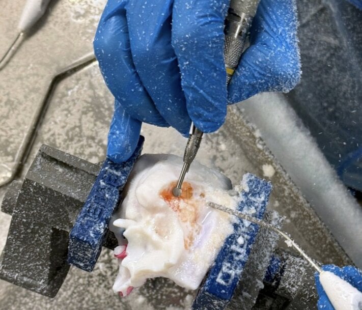
The printed models were designed to respond realistically during surgical procedures. As surgeons drill into the temporal bone, the resistance changes to resemble real tissue, while embedded colour gradients reveal important anatomical landmarks that assist navigation during training.
From geometric accuracy to biomechanical realism
COSA previously produced anatomical models using stereolithography (SLA), which provided high geometric accuracy but could only manufacture parts from a single material. Although suitable for visualisation, these models could not reproduce the layered structure and varying mechanical behaviour of human anatomy.
The adoption of Digital Anatomy technology changed the design process from reproducing geometry alone to engineering both geometry and material behaviour simultaneously.
Medical imaging data is segmented into individual anatomical components, with each structure assigned specific material properties. This enables porous bone, flexible tissues and delicate internal features to respond differently during surgical manipulation.
Developing the final models required extensive optimisation. Material compositions, internal geometries and print parameters were systematically refined through dozens of design iterations, with continuous feedback from practising surgeons to ensure that both tactile response and visual appearance reflected real clinical experience.
Supporting repeatable surgical training
The resulting temporal bone models provide a controlled training environment that overcomes many of the limitations associated with cadaveric specimens.
Instead of relying on whatever anatomy is available, educators can reproduce identical anatomical structures across multiple training sessions. Specific pathologies can also be incorporated intentionally, allowing surgeons to practise targeted procedures under consistent conditions.
This repeatability is particularly valuable for complex interventions such as cochlear implant surgery, where precision and familiarity with anatomical landmarks directly influence surgical performance.
The models also simplify logistics by eliminating the specialised storage, transportation and regulatory requirements associated with cadavers, making advanced surgical simulation more accessible for hospitals, training centres and universities.
Supporting medical device development
Beyond surgical education, COSA's anatomical models are used by medical device manufacturers during preclinical validation and human factors testing.
The consistency of the printed anatomy enables engineers to evaluate cochlear implants, drug delivery systems and other medical devices under repeatable conditions while collecting quantitative data on device-tissue interaction. Identical models also allow multiple clinicians to perform the same procedures, providing more reliable comparisons during usability studies and product development.
Improving efficiency through targeted model design
Although advanced multi-material additive manufacturing involves higher production costs than conventional 3D printing, COSA optimised the manufacturing process by focusing only on the anatomical regions required for training.
Printing only the temporal bone significantly reduces material consumption while preserving the complex internal structures essential for surgical simulation. In addition, PolyJet technology enables multiple models with different anatomical configurations or material combinations to be produced within a single print cycle, improving production efficiency.
Results
By implementing multi-material Digital Anatomy printing, COSA has created high-fidelity temporal bone models that provide a repeatable alternative to cadaver-based surgical training.
The solution enables consistent anatomy across training sessions, realistic tactile and visual feedback during drilling, targeted simulation of specific pathologies, and simplified logistics compared with cadaveric specimens. The same platform also supports preclinical device testing and human factors studies, allowing healthcare organisations and medical technology companies to accelerate surgical training and product development using controlled, reproducible anatomical models.
Edited by Romila DSilva, Induportals Editor, with AI assistance.
www.stratasys.com

